IN MEMORIAM AVINOAM DANIN (1939-2015)
From the website www.shroud.com BARRIE SCHWORTZ

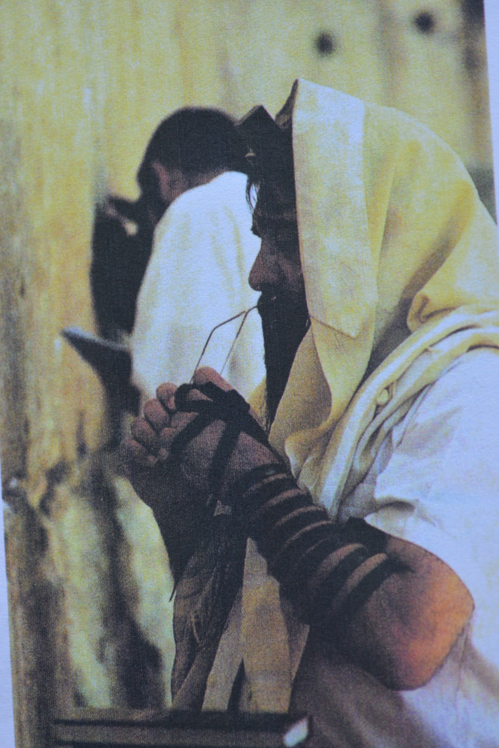
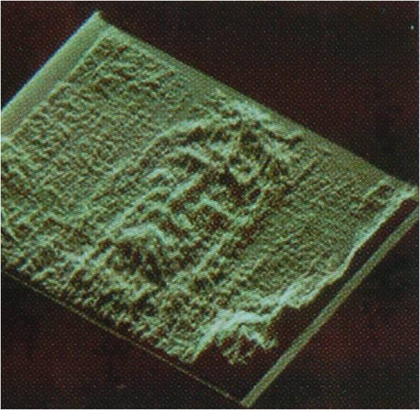
Photo 1. Avinoam Danim
It is with great sadness that I must report the passing of our dear friend and colleague, AVINOAM DANIN, noted Israeli botanist and Shroud researcher, on December 12, 2015, in Jerusalem. The news was first posted on the “Flora of Israel Online” website and I was notified the next day by his friend and colleague, Dr. Anthony Brach of the Harvard University in Cambridge, Massachusetts. Avinoam was well known in the Shroud world for his analysis of the pollens found on the cloth and his claims of finding many flower images on the Shroud. I know that he was frustrated with me at times for not agreeing with some of his imaging claims, but we always enjoyed a respectful and professional relationship and I always considered him a friend. I last saw him in 2013 at the Notre Dame Center in Jerusalem while attending the Third Conference of the Two Linens and we enjoyed a very cordial dialogue. We extend our deepest sympathies and condolences to his family and friends. Rest in peace my friend.
Barrie Schwortz
——————————
Peter Soons was a close friend of Avinoam’s, had met his family in Israel and had worked with him, on a number of research projects. He was kind enough to write a memorial biography for Avinoam and, with the approval of the Danin family, is allowing us to reprint it here in its entirety. We are also including a poignant comment from author Kimberly Ballard who worked with Avinoam during the research and production of her book. The memorial of Dr. Soons:
“For the last 44 years the main academic activity of Avinoam Danin has been dedicated to interdisciplinary research involving the identity and distribution of higher plants, microorganisms, other organisms, and their relation to the environment. The results of his investigations on higher plants and their habitats have been published in 5 books and 182 articles in English, 5 books and 247 articles in Hebrew, one book is bilingual, and one in Italian.
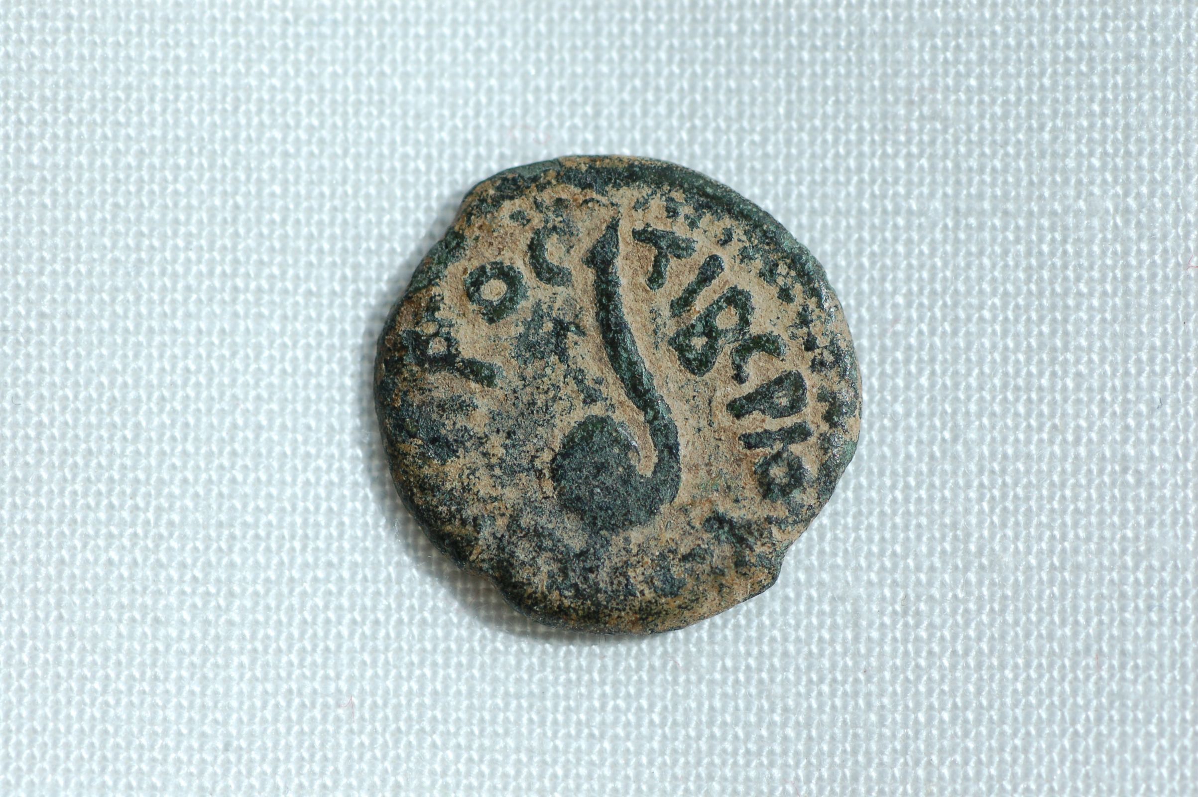
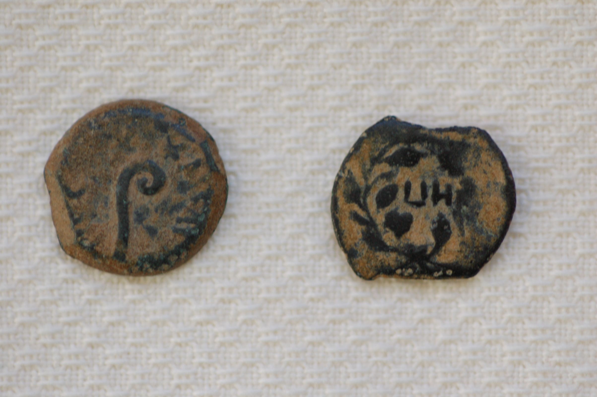
“He discovered many plant species that have not been found previously in Israel, Sinai, and Jordan. In addition, he described more than 40 taxa new to science, belonging to various families. The study of plant names and their use by the Bedouin in the Negev and Sinai led him to cooperate with biochemists and pharmaco-gnosists in the search for natural chemicals, especially in aromatic plants. This contributed to the search for potential medicinal plants. Mapping the distribution of plants in Israel in squares of 5×5 km enabled him to create a data base from which the flora of Israel was studied and a new phytogeographical map was drawn. It was also used in several forensic investigations. The most recent and important one is detecting the origin of the Shroud of Turin in the vicinity of Jerusalem”.
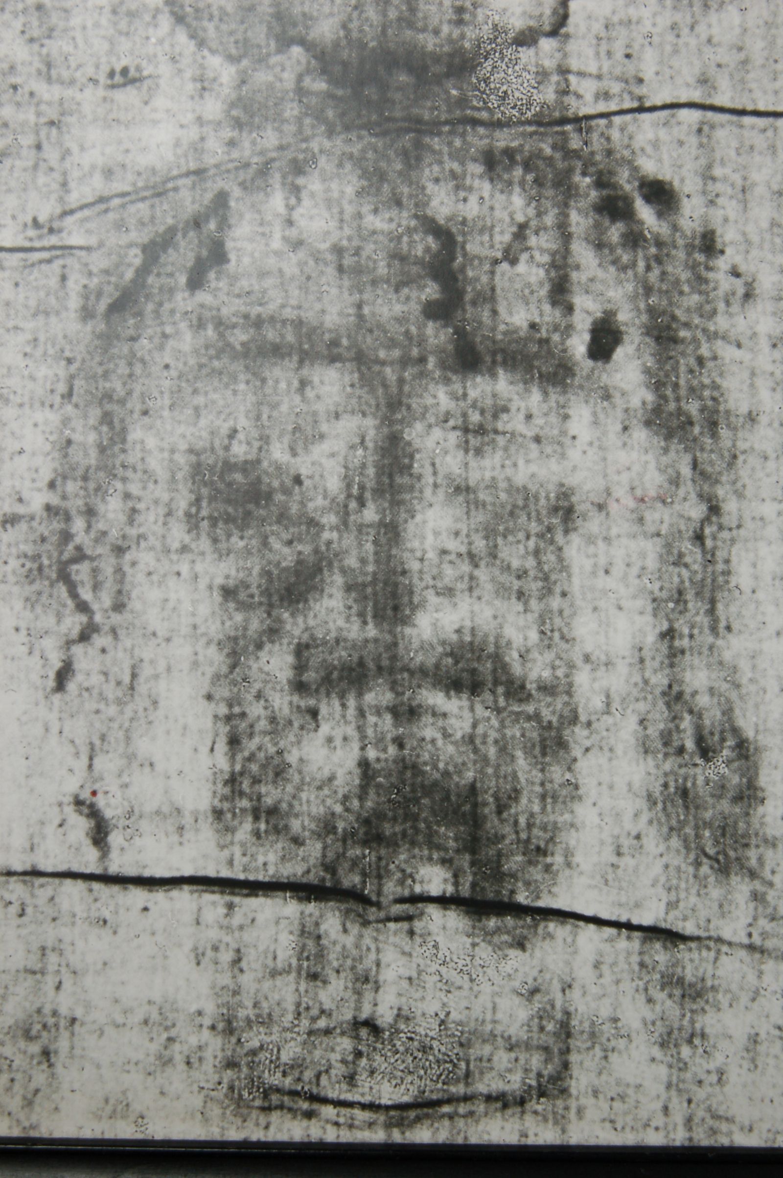
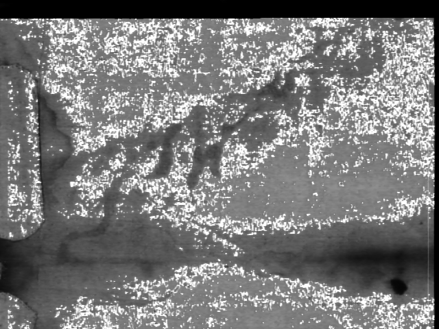
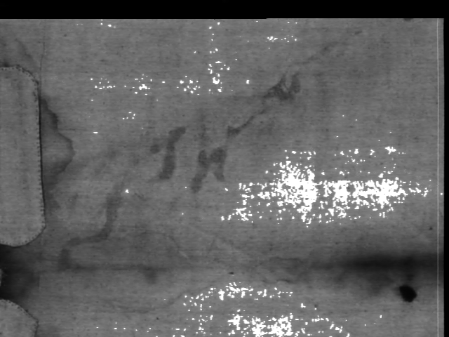
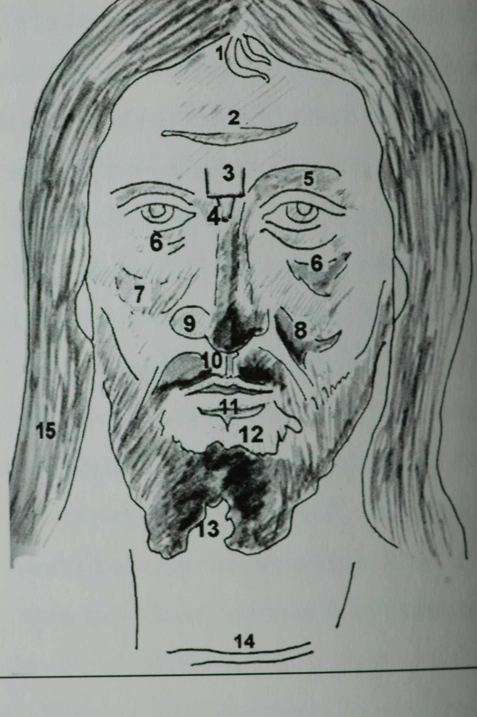
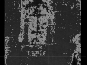
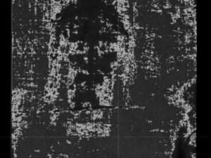
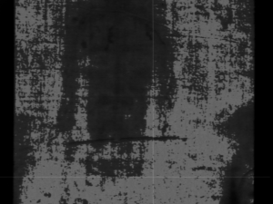
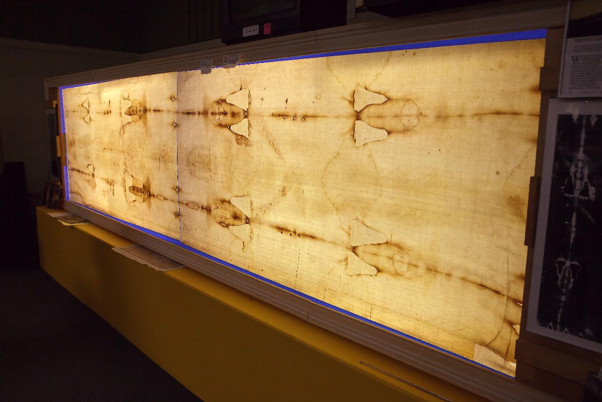
“His involvement with the study of the Shroud started in the early 1980’s when he received a letter from Paul Maloney asking him to do botanical research on the plant images that had been seen on the Shroud. But nothing came of this, until he was visited by Alan and Mary Whanger in September of 1995, who had studied these images. The Whangers determined 28 plant species by comparing the images to 1:1 drawings of plants illustrated in the first three parts of the Flora Palaestina then available. He saw the images they had seen and then found additional ones himself. The conclusions from his botanical research and findings were:
1) The area where the assemblage of three indicator plants could be freshly collected and placed on the Shroud near the man’s body is the area of Jerusalem to Hebron.
2) March-April is the time of the year when the whole assemblage of some ten of the plants identified on the Shroud is in bloom.
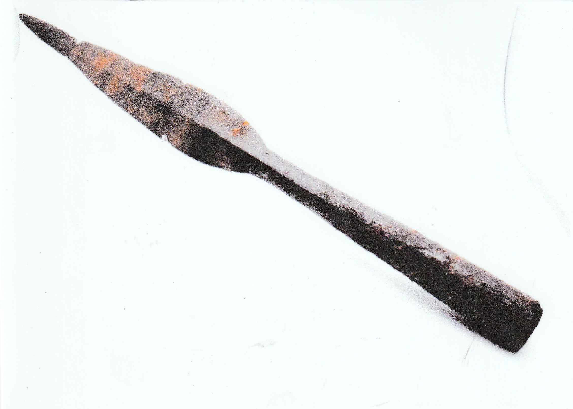
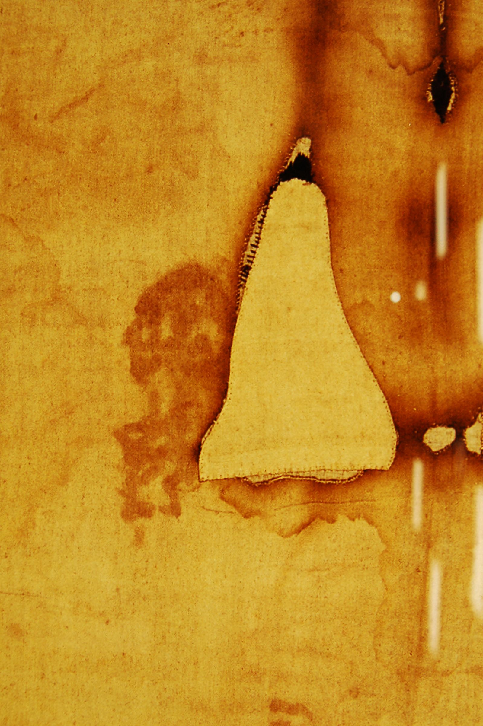
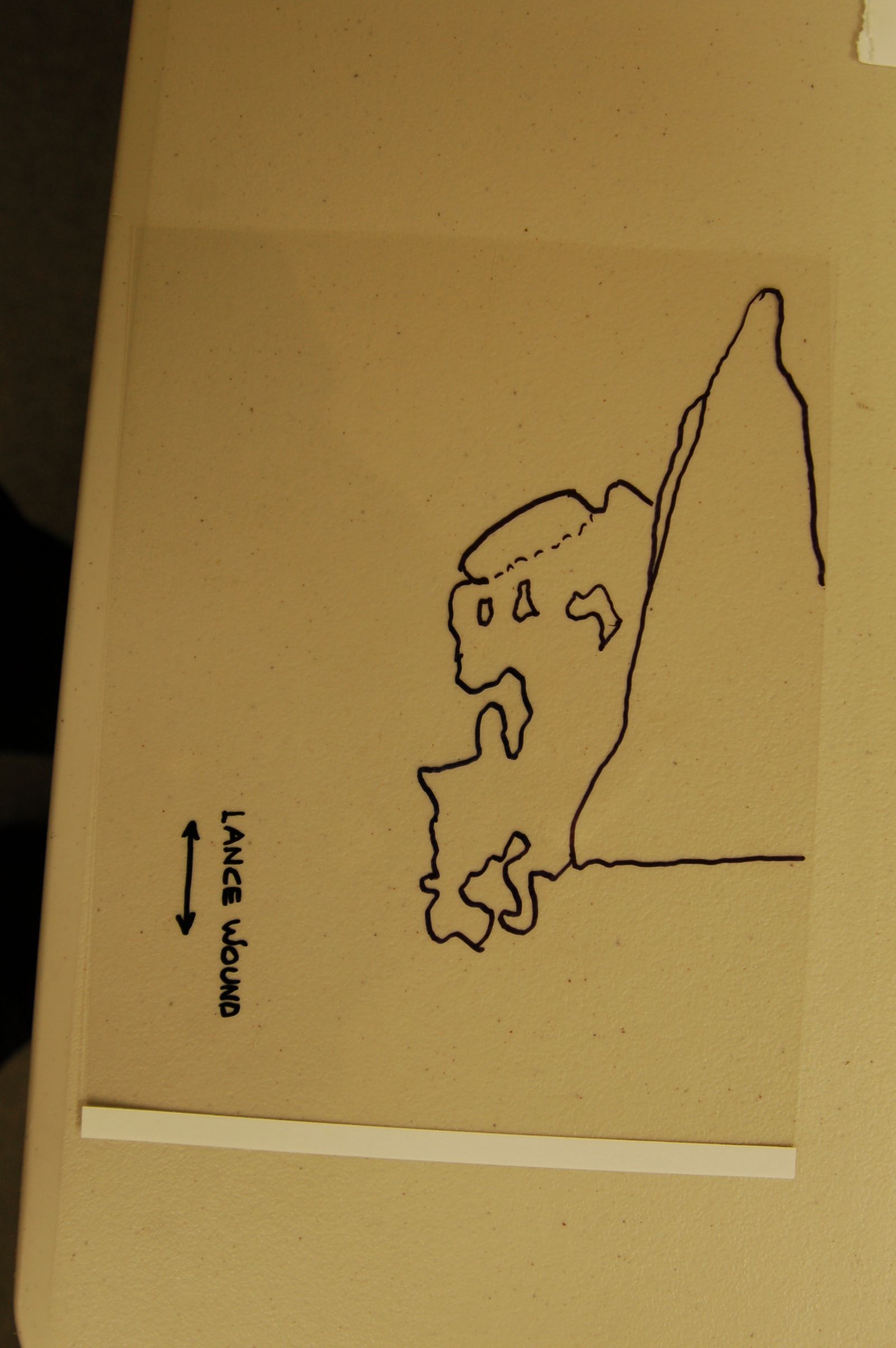
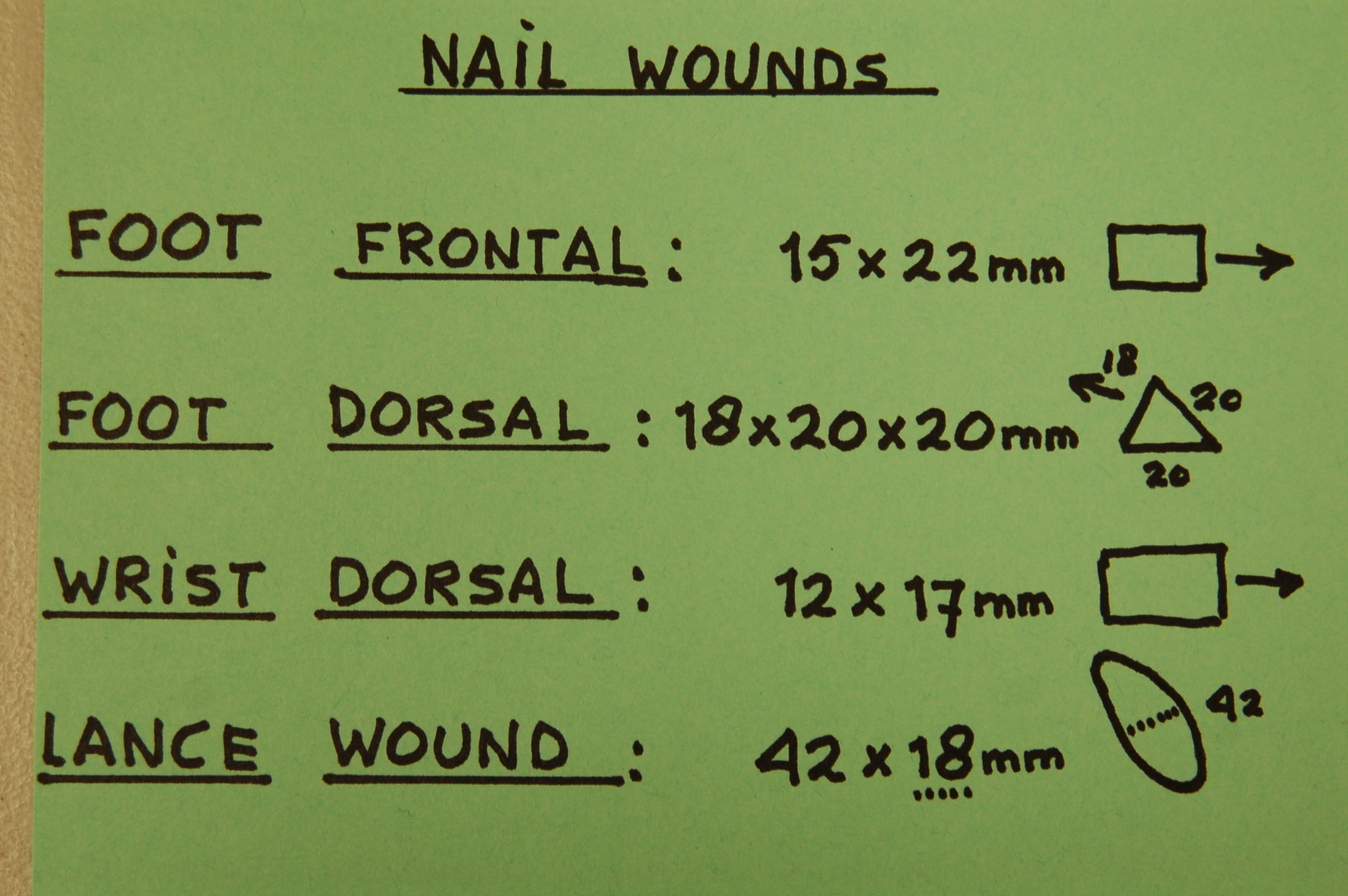
3) Ferocious thorns of two trees from this region were found near the man’s head and a reed was laid alongside his body.
4) Special attention was given to covering parts of the head and face of the Man of the Shroud with daisy-like “flowers” after their carrying stalks were removed.
In 2008, a book interview was done by Father Hector Guerra L.C., that resulted in a book in Italian: “L’Uomo della Sindone”. It was the first book of the series that followed, the English edition: “Botany of the Shroud” (2010), and the Spanish translation: “Flores y Plantas en la Sabana Santa” (2011). (You can see the book cover in the attached photo). Avinoam was also involved in the study of the thorns that were kept in different churches in Italy, and that supposedly had belonged to helmet of thorns of Jesus Christ and to compare them with his findings of the images of thorns on the Shroud.
Using Avinoam’s own words what his investigations of the Shroud meant to him:
“Throughout my years of investigations of the Shroud, I had the pleasure of knowing interesting people and expanding my fields of interest. I lost many hours of sleep as thoughts about the meaning of my findings kept me awake at the oddest hours. I am often asked about my personal feelings concerning the Shroud and what surrounds it. I appreciate the beliefs of Christians around the world and I admire the expression “live and let live”. I have gathered much botanical information in my years of research and am happy to share my expertise and its application in Sindonology with those that are interested”.
Dalys and me will never forget our friendship and all the journeys we made together in Europe and the USA, the presentations we did, our botanical journey in Israel in the springtime to look for the plants you found on the Shroud and the research we did together, but first of all, the joy we had together.
Avinoam, thanks for all your efforts and friendship, also in the name of our sponsor group, and rest in peace.
Peter and Dalys Soons
————————————–
“Avinoam was a light to my world. His kindness to me will never be forgotten. His pictures are forever in my book because he became my friend. I am very sad to lose this good friend. My book is more valuable now because Avinoam’s beautiful pictures are in it. The last time I talked to Avinoam he was having trouble with his feet and feeling a little bit uncomfortable. So, I gave him space to get well. I had hoped that he would come to visit you Barrie and would travel up this way to meet me at some point. I am sad that I will never meet him or be able to ask him anything or show him just how valuable his work was, although I repeatedly told him so. He seems to have doubted his importance. He was a blessing to me. God took him to heaven during Hannukah because he left a legacy of light behind him. I am blessed beyond measure to have had this man as my friend.
Kimberly Ballard
Author


")